How Ultrasound Imaging Helps Doctors Diagnose Health Conditions
Explore the safe, painless, and highly effective world of ultrasound technology and why it's crucial for modern diagnostics.

Mercysam Editorial Board
Medical Content Team

Beyond Pregnancy: The Power of Sonography
When most people hear the word "ultrasound," they immediately think of obstetrics and viewing a developing baby. While it is an indispensable tool for monitoring pregnancy, ultrasound technology (sonography) is a highly versatile diagnostic tool used across nearly every medical discipline to peer inside the human body safely and in real-time.
How Ultrasound Works
Unlike X-rays or CT scans, which use ionizing radiation, ultrasound relies on high-frequency sound waves. A handheld device called a transducer is moved over the skin (often with a special gel). The transducer emits sound waves that bounce off internal organs, tissues, and fluids. The returning echoes are captured and instantly translated by a computer into real-time visual images on a monitor.
Diagnosing Abdominal and Pelvic Issues
Ultrasound is frequently the first line of investigation for abdominal pain. It provides excellent visualization of solid organs like the liver, gallbladder, spleen, pancreas, and kidneys. It is highly effective at detecting gallstones, kidney stones, liver disease, and abdominal masses. In the pelvic region, it is crucial for evaluating the uterus and ovaries, helping to diagnose conditions like fibroids, cysts, and endometriosis.
Cardiovascular Applications (Echocardiogram)
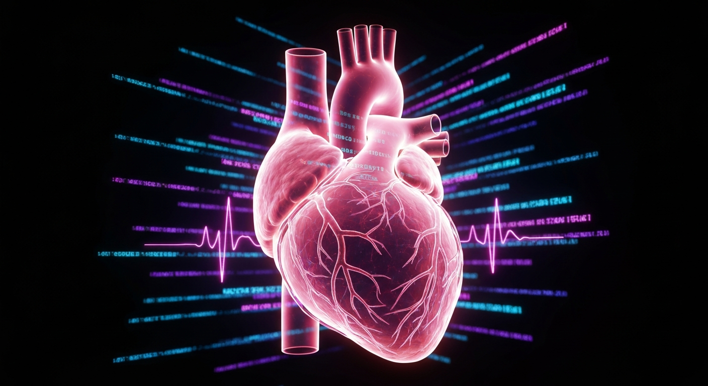
When an ultrasound is performed on the heart, it's called an echocardiogram. This specialized test allows cardiologists to see the heart beating and pumping blood in real-time. It can identify abnormalities in the heart valves, measure the strength of the heart muscle, and detect fluid buildup around the heart, making it vital for diagnosing heart failure and structural heart disease.
The Safety Profile of Ultrasound
One of the greatest advantages of ultrasound imaging is its safety profile. Because it uses sound waves rather than radiation, it is considered completely safe for pregnant women, children, and adults requiring frequent monitoring. It is non-invasive, painless, and typically requires no special preparation, making it an incredibly patient-friendly procedure.
Real-Time Dynamic Imaging
Unlike static images produced by X-rays, ultrasound provides dynamic, real-time imaging. This allows doctors to observe blood flowing through arteries and veins (using Doppler ultrasound), watch a tendon glide over a joint, or guide a needle precisely during a biopsy. This real-time capability is invaluable for accurate, targeted medical interventions.
Conclusion: A Pillar of Modern Medicine
Ultrasound is a fast, safe, and incredibly informative diagnostic tool. At Mercysam Diagnostic Centre, we utilize advanced 3D and 4D ultrasound equipment operated by highly skilled sonographers to ensure the clearest images and the most accurate diagnoses possible, aiding your doctor in providing the best care.